Clinical research is a cornerstone of modern medicine, driving discoveries that improve treatments, understand diseases, and save lives. Yet despite technological advances, traditional clinical trials are often slow, expensive, and labour-intensive. Collecting and analysing large volumes of data, from patient records to imaging studies, can take months or even years.
Artificial intelligence (AI) is now transforming this landscape, providing tools that speed up research, reduce costs, and improve accuracy. Among the most promising applications is medical image analysis, where AI can automate or assist with tasks that were once boring and time-consuming. One example comes from MIT researchers who developed a system called MultiverSeg to improve the segmentation of medical images [1].
The Challenge of Medical Image Segmentation
In many clinical studies, researchers begin by identifying regions of interest in biomedical images—a process called segmentation. For instance, neuroscientists studying memory-related diseases may want to measure changes in the hippocampus, a small brain structure crucial for memory. To do this, each hippocampus must be outlined in every brain scan in the dataset.
For large studies, this can be daunting. Some research datasets include hundreds or even thousands of images. Manually outlining each structure can take hours per image, and the task becomes even more difficult when the anatomy is small, irregular, or affected by disease.
Traditional AI approaches have tried to automate segmentation. One method is interactive segmentation, where a researcher marks parts of an image and the AI predicts the rest. Another approach involves training a task-specific AI model, which requires manually segmenting hundreds of images to create a labeled dataset. Both approaches have limitations: interactive systems require repeated effort for every image, while task-specific models need extensive training and cannot easily adapt to new data or corrections.
MultiverSeg: A Smarter Approach
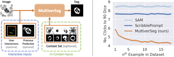
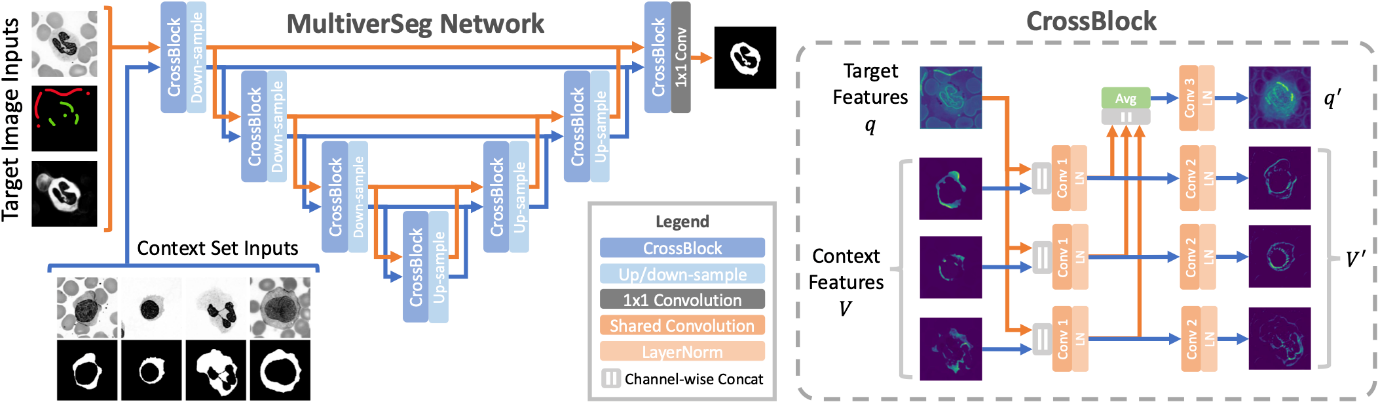
MultiverSeg combines the best of both worlds. It is an AI system that can learn from user interactions while also referencing previously segmented images to make more accurate predictions.
The process is simple and intuitive: researchers upload an image, mark regions of interest with clicks, scribbles, or boxes, and the AI predicts the full segmentation. The more images the researcher annotates, the better the AI becomes, eventually reaching a point where it can segment new images without any additional input.
Unlike traditional task-specific models, MultiverSeg does not require pre-labeled training datasets. This means that researchers without machine-learning expertise or large computational resources can use the system immediately for new studies. The model’s context-aware architecture allows it to refer to a growing “context set” of previously segmented images, improving predictions over time and reducing the effort required from the user.
In comparative studies, MultiverSeg outperformed existing state-of-the-art segmentation tools. By the ninth new image, it required only two user clicks to achieve higher accuracy than models designed specifically for the task. For simpler image types, like X-rays, only one or two manually segmented images are sufficient for the AI to perform independently.
Impact on Clinical Research
The implications of such technology are profound. Manual image segmentation has long been a bottleneck in clinical research. Many scientists might only manage to segment a few images per day, limiting the size and scope of their studies. MultiverSeg dramatically reduces this burden, enabling researchers to focus on analysis and discovery rather than repetitive annotation.
By accelerating segmentation, AI tools like MultiverSeg could also reduce the cost and duration of clinical trials. Faster data analysis means quicker insights into disease progression, treatment efficacy, and patient outcomes. This efficiency could be particularly valuable in urgent medical contexts, such as evaluating treatments for emerging diseases or personalizing therapies based on patient-specific imaging.
Moreover, the tool has potential applications in clinical practice. For example, radiation oncologists could use AI-assisted segmentation to plan treatment more accurately and efficiently. By reducing manual workload, physicians could spend more time on patient care and critical decision-making.
User-Centered AI Design
One of the reasons MultiverSeg is so promising is its interactivity and flexibility. Researchers can iteratively correct AI predictions, refining results to the desired level of accuracy. The system is also designed to handle datasets of any size, making it adaptable to different studies, imaging modalities, and clinical requirements.
Hallee Wong, the lead author of the study, emphasizes that the tool “enables new science by allowing clinical researchers to conduct studies they were previously unable to do due to the lack of an efficient tool.” By reducing the friction of segmentation, AI not only saves time but also empowers scientists to explore research questions that were previously impractical.
Future Directions
Looking forward, the research team plans to test MultiverSeg in real-world clinical settings and gather feedback from collaborators to improve usability. They also aim to expand the tool’s capabilities to 3D biomedical images, which are increasingly common in advanced imaging studies such as MRI and CT scans.
The broader vision is clear: AI tools that streamline repetitive or labor-intensive tasks will allow clinical researchers and physicians to focus on analysis, interpretation, and decision-making. By integrating AI into the workflow, medical studies can be conducted faster, with fewer errors, and at a lower cost, ultimately accelerating the pace of medical innovation.
References: 1) Adam Zewe, MIT News. 2) Wong, H., et al., MultiverSeg: Scalable Interactive Segmentation of Biomedical Imaging Datasets with In-Context Guidance (2024). 3) Sébastien Goll et al., MultiverSeg segment editor effect. 4) Butoi, V., et al., UniverSeg: Universal Medical Image Segmentation (2023).